


Characterization of biological materials
Characterization of biological materials
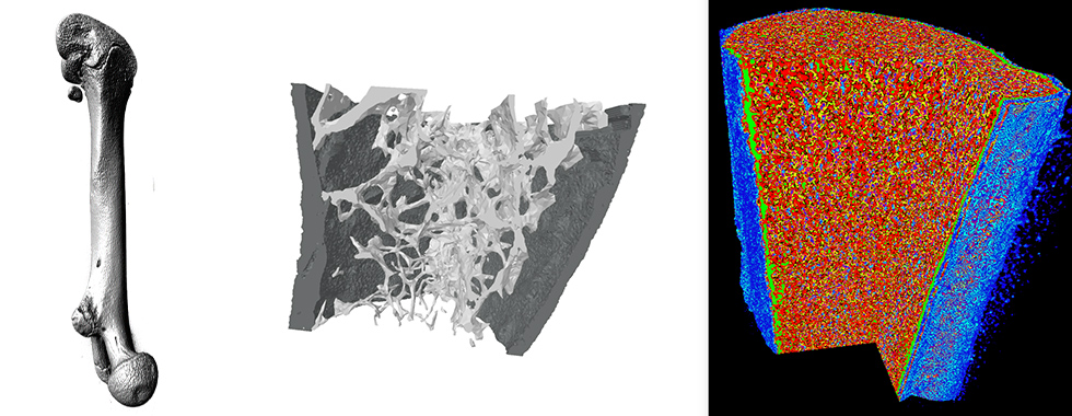
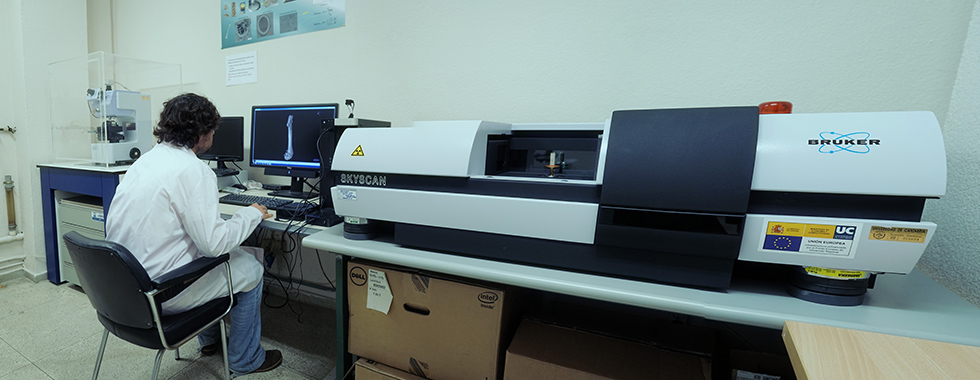
Micro-tomografía axial computerizada (μtac)
LADICIM has a Bruke (model Skyscan 1172) computerized axial micro-tomograph (μCT). The most relevant technical characteristics are: X-ray camera of 11 Mp with correction of total distortion, up to 8000 × 8000 pixels in each layer, isotropic detail detectability up to 0.7 μm and software for 2D and 3D image analysis. The main bone parameters that the μTAC is able to identify are the following: porosity, cortical volume, trabecular volume, connectivity, degree of anisotropy, etc.
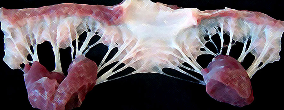
The figure on the left shows the microstructure of a functional mitral tendon cord. The interior configuration of the collagen fibres is appreciated, in an undulating arrangement, covered with several layers of elastin and the outer region of endothelial cells. The figure on the right shows a calcified mitral cord. The bright granules that are observed in the upper part correspond to calcium accumulations.
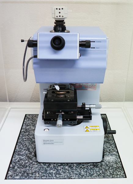
Ultra-microindentation (UMI)
The UMI tests are carried out with a Shimadzu DUH 211, Dynamic Ultra-Micro Hardness Tester, equipped with a Berkovich tip. The load capacity goes from 0.1 mN to 1961 mN. This test allows the hardness of the material (in different scales: Vickers, Martens, etc.) to be determined and provides estimations, at local scale, of relevant mechanical parameters, such as the Young’s modulus of the material.
Mechanical tests
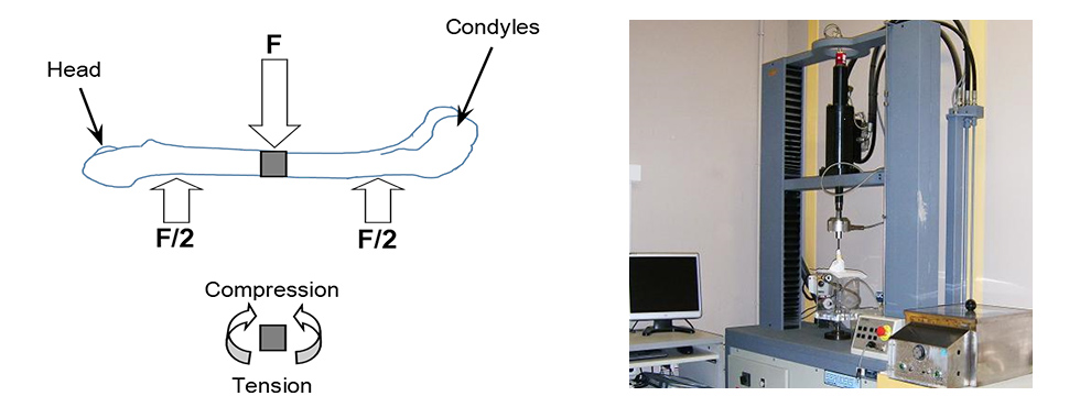
The bending tests allow overall resistant properties of the bones such as the force at fracture, the force corresponding to yield strength, the deformation at fracture or the energy absorbed up to the maximum load or to the breaking load to be obtained. It is possible to carry out bending tests in three and four points. Once the resistant cross section of the bone is determined by means of μCT, intrinsic resistant results of the material can be derived, such as the Young’s modulus or the yield strength. The bending tests are carried out by means of a Servosis electromechanical machine, model ME-402. This machine is equipped with a low capacity load cell, specifically designed for testing small bones. The tests are carried out under physiological conditions, in an urn equipped with a serum recirculation system at 37ºC.
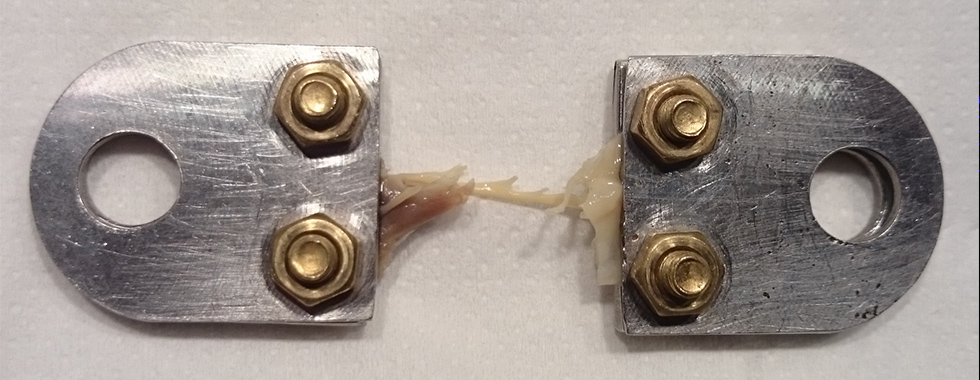
The tensile tests allow the global resistant properties of the biological tissues such as the fracture strength, the force corresponding to the yield strength, the elongation at fracture or the energy absorbed up to maximum load or to the breaking load to be obtained. Once the resistant cross section of the element (for example, a chordae tendineae or an artery) is determined by means of μCT, intrinsic resistant results of the material can be derived, such as its Young’s modulus or its yield strength. The tests are carried out in a Servosis electromechanical machine, model ME-402. This machine is equipped with a low capacity load cell, specifically designed for testing small bones. The tests are carried out under physiological conditions, in an urn equipped with a serum recirculation system at 37ºC. Using the same equipment, it is possible to perform fatigue tests in which the component is subjected to variable loads over time, simulating the effect caused by the heartbeat.
Scanning Electron Microscopy (SEM)
Zeiss Scanning Electron Microscope (SEM) model EVO MA 15. This equipment can work both in high vacuum and in atmospheric conditions allowing images on non-conductive materials to be acquired. The electron acceleration voltage reaches 30 kV and the magnification varies between x7 and x106. It has a microanalysis system by dispersive energy spectroscopy (EDS) that allows the chemical nature of the materials analyzed to be determined.
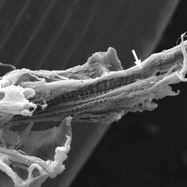
The figure shows a micrograph obtained by SEM of a mitral tendon cord subjected to a tensile test, stopped when detecting a load drop. The SEM examination allowed the micro-fractures in the collagen fibers, responsible for the reduction in the resistant capacity of the component, to be located.

In vitro tests and animal models
In collaboration with the Bone Genomics Laboratory (IDIVAL-UC), several in vitro tests and animal models are performed, including cell cultures, genotyping and gene expression analysis, histological studies, characterization of drug effects and fracture models.